Featured Scientist: Kiley Hughes, PhD candidate (Anticipated Spring 2022), School of Biological Sciences, Illinois State University

Birthplace: Seattle, WA
My Research: I study how Duchenne muscular dystrophy progresses. My research is focused on figuring out how the absence of the dystrophin protein causes the disease.
Research Goals: I want to continue using behavioral and molecular techniques to research neuromuscular disorders.
Career Goals: I want to be a scientist! I would love to work on a larger research team for a research institute that shares their work with the public.
Hobbies: I like to cook and paint, go outside when the weather allows it, and play with my two cats.
Favorite Thing About Science: My favorite thing about science is the unknown, there is always more to know and understand. I allow my curiosity to drive me.
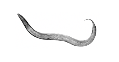
Organism of Study: I study a species of microscopic worm, Caenorhabditis elegans. C. elegans is a model organism, which means that scientists have been using it for research for many years and we know a lot about it. This species of worm is often used to study human diseases.

Field of Study: Molecular Neuroethology
What is Molecular Neuroethology? In this field, we use molecular techniques to understand the way organisms interact with the environment and specifically look at the cause of different types of behavior.
Check Out My Original Paper: “Physical exertion exacerbates decline in the musculature of an animal model of Duchenne muscular dystrophy”

Citation: K.J. Hughes, A. Rodriguez, K.M. Flatt, S. Ray, A. Schuler, B. Rodemoyer, V. Veerappan, K. Cuciarone, A. Kullman, C. Lim, N. Gutta, Physical exertion exacerbates decline in the musculature of an animal model of Duchenne muscular dystrophy. PNAS. 116.9: 3508-3517 (2019).
Research at a Glance: Duchenne muscular dystrophy is a degenerative disease that affects 1 in 3500 males and there is still a lot that we do not know about this disease. My research looks at how the loss of the muscle protein, dystrophin, leads to muscle death. I study the extent to which exercise might be able to protect dystrophic muscles. In this study, we used the nematode worm, C. elegans, and our worms have Duchenne muscular dystrophy. We used them to show that calcium was not being properly regulated in their bodies, even before the worms show symptoms of the disease. We also observed that some types of muscle activity increased muscle repair. However, no treatment positively affected the life expectancy of dystrophic animals.
Highlights: For a muscle to contract, the body needs both energy and calcium. We used a special worm that has a green protein that will glow when it is in the presence of calcium. Worms move in a wave-like fashion. Because of this, each contracting muscle should have an opposite relaxed muscle. We used our special worm to do in vivo calcium measurements (Figure 1).

We found that in dystrophic worms, not only was muscle calcium higher, but this persisted in both contracting and relaxing muscles.
What My Science Looks Like: We can use electron micrographs to see what happens to the muscles in worms that have Duchenne muscular dystrophy. We make them by taking a worm and freezing it under very high pressure. The frozen worms are treated with heavy metals, then cut into really small slices (like pastrami). The slices shown in Figure 2 are from the midbody of a worm and they show the worm’s muscle structure.

The image on the left is from a wild type worm, which means that this worm does not have the disease. The left image shows what a normal, healthy, muscle should look like. The image on the right shows the muscle structure of a dystrophic worm. The muscle in the dystrophic worm is degenerating.
The Big Picture: Thousands of people are living with Duchenne muscular dystrophy in the United States. These patients are wheelchair bound by the age of 12 and die prematurely from heart failure in their 30’s. Research has been slow in this field because most of the animals that have the human version of Duchenne muscular dystrophy do not show the same type of degeneration. However, when our dystrophic worms burrow, they show muscle decline similar to humans. Using this model, we hope to understand how people with this disease are getting so sick.
Decoding the Language:
Caenorhabditis elegans (C. elegans): The worms that we use to study Duchenne muscular dystrophy. This might seem strange, but they’re a great model organism to understand the disease. What we find can be translated to humans!
Degenerative disease: Diseases that are caused by the abnormal break down of cells over time.
Duchenne Muscular Dystrophy: A fatal neuromuscular disorder that causes weakness in and loss of skeletal and heart muscle. This disease is caused by abnormal dystrophin proteins.
Dystrophic: A term used to refer to animals that have absent or abnormal dystrophin proteins.
Dystrophin: The muscle gene/protein that is absent in patients with Duchenne muscular dystrophy.
Electron micrograph: A form of imaging that can be done using a specialized microscope.
In vivo: Measurements or experimental procedures that take place inside of a living organism.
Microbes: A microscopic organism, like bacteria.
Model organism: A species that has been very widely studied. These species are often easy to breed and to take care of in the lab. Because they have been studied for so long, scientists have developed a lot of genetic resources that can be used to model human diseases using these animals.
Neuromuscular disorders: These are diseases that affect muscles and how the nervous system controls those muscles. Molecular techniques: A method used to manipulate and understand DNA, RNA, or protein.
Wild type: An animal that has not been manipulated. It should have the same genetic and physical characteristics as if it were found in the wild.
Learn More:
Why use the worm?
Synopsis edited by: Eric Walsh, M.S. 2020, and Rosario Marroquin- Flores, PhD (Anticipated Spring 2022), School of Biological Sciences, Illinois State University.
Download this article here
Please take a survey to share your thoughts about the article!
