Featured Scientist: Mark A. Rossi, Ph.D., Psychiatry Department, Rutgers Robert Wood Johnson Medical School, Child Health Institute of New Jersey.

Birthplace: Detroit, Michigan.
My Research: I attempt to understand what motivates our behavior and the way that we eat. I focus on how the brain is wired and I am particularly interested in how our diet can rewire our brain circuits.
Research Goals : My goal is to help develop therapies that can treat eating disorders, like diabetes.
Career Goals: I want to push the technological boundaries of how we study the brain.
Hobbies: Soccer! I’ve played all my life.
Favorite Thing About Science: The freedom and flexibility to go where the data takes me.
My Team: I recently opened my own lab, so I am temporarily a team of 1. The lab will soon be populated with research technicians, graduate students, postdoctoral researchers, and undergraduates.
Organism of Study: Mice

Field of Study: Neuroscience
What is Neuroscience: Neuroscience is the study of the nervous system. The goal of this field is to understand how the central nervous system works. The central nervous system includes the brain and the spinal cord. My area of expertise is in how the brain contributes to motivated behavior. Motivated behavior includes any type of behavior that is important for our survival.
Check Out My Original Paper: “Obesity remodels activity and transcriptional state of a lateral hypothalamic brake on feeding”

Citation: Rossi MA, Basiri ML, McHenry JA, Kosyk O, Otis JM, van den Munkhof H, Bryois J, Hubel C, Breen G, Guo W, Bulik CM, Sullivan PF & Stuber GD. (2019) Obesity remodels activity and transcriptional state of a lateral hypothalamic brake on feeding. Science. 364(6447):1271-1274.
Research At A Glance: Obesity is an inflammatory condition where our bodies store extra fat. Obesity can increase our chances of developing health issues, such as heart disease. I am mainly interested in understanding the role that our brain plays in obesity. A region of the brain called the hypothalamus is responsible for coordinating many day-to-day actions such as, reproduction, aggression, and feeding. The lateral hypothalamic area (LHA) is an area within the hypothalamus that has neurons that are known to control feeding behavior. A neuron is a brain cell that can communicate with other cells using electrical signals. We don’t know very much about the neurons in the LHA, and it is not clear if they are affected by obesity. In our paper, we characterize the excitatory neurons in the LHA and try to understand how they may be affected by obesity. We used a technique called single-cell sequencing to look at the messenger RNA (mRNA) in a single cell and compare it to other individual cells. This allows us to build a profile of the cells in LHA. In our experiments, we looked at the brain to find differences between lean and obese mice. We found that the pattern of gene expression was different between the mice, particularly within the excitatory neurons. Excitatory neurons are a type of neuron that increase the likelihood that a neuron will create and fire an action potential. In normal conditions, the excitatory neurons will send signals to activate other neurons that function as a ‘brake’ to suppress food intake. We found that a high fat diet can change the activity of excitatory neurons, so they no longer send the signal to suppress food intake. Next, we wanted to see if the cells change in function in response to obesity. We used a technique called longitudinal calcium imaging to magnify and take pictures of the cells under a microscope over the course of 12 weeks. We found that the normal functions of excitatory neurons in the LHA were greatly decreased in obese mice. Our research gives us one mechanism for how the brain may be altered by diet. As we become obese, the excitatory neurons in our brains have reduced activity, which may contribute to the behavior of overeating.
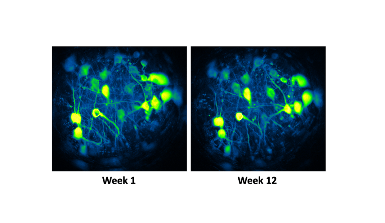
Highlights: One of the goals of our study was to see how the activity of the LHA excitatory neurons change in mice with a high fat diet and how this may contribute to obesity. What we found is that a chronic high fat diet affects how excitatory neurons function,and this can increase overeating. The most important part of this project was our ability to track the activity of individual neurons in the hypothalamus over time (Figure 1). We monitored the mice as they started the high fat diet and as they became obese. This helped us show that while the structure of the individual neurons looked similar in the brain, they did change in function in response to diet over time. That is, single neurons will respond to food rewards but lose that responsiveness as obesity progresses.

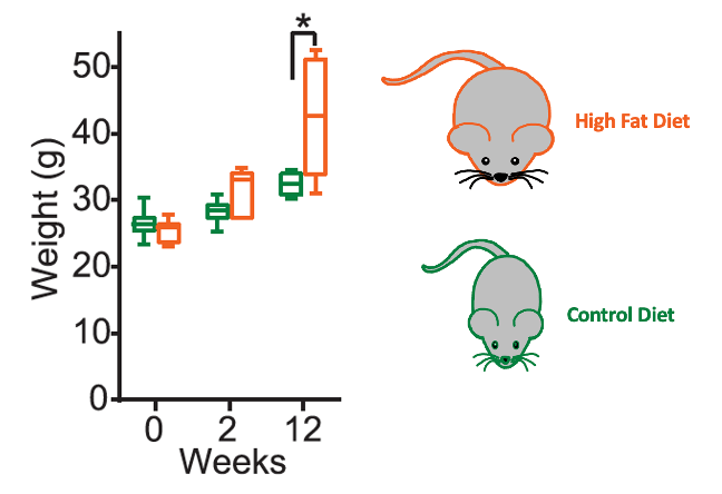
What My Science Looks Like: For this experiment, we fed six mice (n=6) a control diet and seven mice (n=7) a high fat diet for 12 weeks (Figure 2). The high fat diet had higher levels of fat content because it had high levels of sucrose (sugar). Like humans, a diet with a high fat content leads to an increase in body weight for mice. After 12 weeks, mice fed with the high fat diet gain significantly more weight than mice fed on the control diet (Figure 2).
The Big Picture: Obesity is a serious medical problem that is widespread in the United States. It is associated with increased risk of death from heart disease, stroke, and diabetes. While it affects many people, there are few viable treatment options. It is important to understand how the brain controls normal eating and overeating. This knowledge is critical to help us develop new treatments for eating disorders and obesity. Our research tries to understand how the brain contributes to normal feeding. It also looks at how those same brain circuits are affected by an unhealthy diet. Measuring the activity of neurons in the hypothalamus using multiple methods can help us get a more complete picture of the ways that the brain controls feeding behavior. Our research provides us with important insights and a deeper understanding of how the brain can be changed by obesity.
Decoding the language:
Action potential: Action potentials are electrical signals that allow information to be transferred to the nervous system. The nervous system will then transmit information to its target cells.
Brain circuits: Brain circuits consist of a web of neurons that are connected to each other. Information will flow from one neuron to another to send information to other parts of the brain. This is similar to the electrical circuits in our homes. When you walk into a room and flip a light switch, the electrical current will be carried across the room to turn on a light bulb.
Neuron: A neuron is a cell that acts as messenger. It sends and receives information to and from the brain and spinal cord. This information then goes to different areas of the body. For example, when you are hungry your stomach will send a signal to a region in your brain called the hypothalamus to let you know that you need food!
Excitatory neurons: Excitatory neurons are cells in the brain that increase the likelihood that a neuron will create and fire an action potential. When neurons fire an action potential, they are using electrical signals to communicate to other cells. The excitatory neurons are distinct from other types of neurons, like inhibitory neurons, because they increase the signal, rather than silencing it.
Gene expression: Gene expression allows us to measure gene activity. At any given time, our bodies will turn on genes to perform important bodily functions. For example, if you are eating a sandwich, your body may turn on a gene that codes for an enzyme to help break down the food in your mouth. Before a gene can perform a function, it must be converted into messenger RNA (mRNA). Therefore, we can measure the expression of a specific gene by quantifying how much mRNA for that gene is in a sample. We say that a gene has high expression when the mRNA for that gene is very abundant in our sample. In our study, we changed the diets of our mice and measured which genes had high expression after the change in diet. This helps us to understand the role of certain genes in conditions like obesity.
Genetically encoded fluorescent calcium indicator (GECI): GECI is a tool that marks a set of neuron cells by binding to calcium ions. This allows us to track the calcium activity which is associated with the firing of action potentials. Higher calcium activity will manifest as a brighter signal, with the help of a fluorescent dye. It allows us to read the activity of the neurons to better understand how neuronal activity relates to behavior.
Hypothalamus: The hypothalamusis an area in the brain that plays an essential role in regulating motivated behaviors. This can include hunger, reward responses, and homeostasis. The hypothalamushelps to keep the body stable and plays a role in heart rate and blood pressure.
Lateral Hypothalamus area (LHA): The LHA is a small region in the hypothalamus that regulates feeding behavior. In this research, we looked at cells in the LHA to see how these cells were affected by obesity.
Messenger RNA (mRNA): mRNA are intermediate molecules between DNA and proteins. In our research, we used mRNA to look at patterns of gene expression. This allowed us to identify which genes were being activated in the LHA neurons in response to our control and high fat diet.
Single-cell sequencing: Single cell sequencingis a specific form of gene sequencing. Gene sequencing allows us to read the genetic code in DNA and quantify how much of each gene is present in a sample. Most gene sequencing techniques read the genetic code of several cells at one time. The single cell sequencing method is unique because it allows us to measure gene expression in individual cells. This method allowed us to target excitatory neurons and see how diet impacts gene expression in these specific cells.
Learn More:
Video on how neurons communicate
More research papers about how the circuits in the brain contribute to feeding behavior:
Rossi MA & Stuber GD (2018) Overlapping brain circuits for homeostatic and hedonic feeding. Cell Metabolism, 27(1): 42-56.
Rossi MA, Marcus L. Basiri, Yuejia Liu, …, Charu Ramakrishnan, Karl Deisseroth, and Garret D. Stuber (2021). Transcriptional and functional divergence in lateral hypothalamic glutamate neurons projecting to the lateral habenula and ventral tegmental area. Neuron, 109, 1-15.
Synopsis edited by Maisam Yousef, B.S. 2019, Illinois State University, and Naiomy Rios Arce, PhD.
Download this article here
Please take a survey to share your thoughts about the article!
