Featured Scientist: Dr. Thomas Longden (he/him/his), Assistant Professor, University of Maryland School of Medicine, Department of Pharmacology, Physiology and Drug Development

My Research: My work focuses on developing our understanding of how blood flow is controlled in the brain. My work also focuses on how blood flow supports the constantly changing energy needs of our brain cells.
Research Goals: My future research goals include building a complete picture of how brain cell activity controls blood flow. I would also like to determine whether protecting or rescuing blood flow in the brain halts dementia and other diseases of the aging brain.
Career Goals: What I am doing now! My goal has always been to run my own research group and work on problems I find interesting and that I think might benefit others. I hope to be able to continue to do this and expand my research group so we can do more and more impactful science.
Hobbies: Rock climbing and bird watching.
Favorite Thing About Science: My favorite thing about science is the feeling of being the first and only person in history to know a particular something about the universe. The feeling after a successful new experiment is particularly special. I don’t think you get this in other domains, and I have been chasing this feeling since my first experiment!
Scientist Upbringing: My parents were both teachers, and my dad taught chemistry and physics, so my act of rebellion was to study biology. I first wanted to become a scientist when I started learning about evolution, which was a big ‘aha’ moment for me and answered a lot of questions I had about the world at that time. From there I became more and more interested in biological systems – how they work and how they came to be. That interest took me to the University of Manchester for my undergraduate degree in Pharmacology, and there I also completed a PhD under Gill Edwards and Arthur Weston. During my PhD work, I spent time in Germany and in the U.S. in the lab of Mark Nelson, who then became my postdoctoral mentor.
My Team: I work with an amazing group of scientists! Our team currently numbers four graduate students and two postdoctoral researchers. Each person is working on distinct exciting projects, but we ensure there is a good amount of overlap of skills and interests which helps us to make use of a wide range of techniques to address our work.
This project was the last work I did with Dr. Mark Nelson before starting my own lab, so I made most of the recordings and did a lot of the analysis and writing. I was also accompanied by Dr. Grant Hennig, who wrote thousands of lines of code to enable the data analysis, and Dr. Amreen Mughal, who also played a key role in acquiring data. I remember this project very fondly – lots of days sitting at the microscope watching cells light up and chatter among themselves in mysterious patterns!
Organism of Study: Mouse, primarily. Because the biology of mice is very similar to that of humans, they are often bred specifically for research, and then used to better understand how we may improve human health.Mice allow us to explore biological systems in ways that wouldn’t be feasible in humans. Recently, we’ve been moving towards working with human cells.

Field of Study: Physiology
What is Physiology? Physiology is the study of how systems and processes work within a living organism. These might occur within the cell, between cells, within an organ, or between organs. For example, researchers might do physiology studies to understand how the heartworks with the blood vessels to deliver nutrients to all the cells and organs in the body so that they can carry out the processes which keep you alive.
Check Out My Original Article: “Local IP3 receptor–mediated Ca2+ signals compound to direct blood flow in brain capillaries”

Citation: Longden TA, Mughal A, Hennig GW, Harraz OF, Shui B, Lee FK, Lee JC, Reining S, Kotlikoff MI, König GM, Kostenis E, Hill-Eubanks D, Nelson MT. (2021). Local IP3 receptor-mediated Ca2+ signals compound to direct blood flow in brain capillaries. Science Advances, 7(30). https://doi.org/10.1126%2Fsciadv.abh0101.
Article written by Dr. Jennifer McFarland (she/her), University of Maryland School of Medicine.
Research At A Glance: When we think, understand, sense things, and move, our brain cells become more active. Our brain cells also regulate processes that we don’t think about, like controlling our heartbeat. Highly active brain cells need extra energy. Blood cells carry nutrients to the cells in our bodies. Our blood cells cantransport and provide energy to active brain cells in the form of oxygen, and travel along with the sugar our brains need through a network of tubes called blood vessels. The walls of our blood vessels are made up of two types of cells. Endothelial cells form the barrier between the blood and the rest of the body, allowing the blood to be contained. These cells are surrounded by another type of cell called pericytes. Some pericytes constrict and relax, and this allows the walls of the blood vessels to narrow or widen. Blood vessels allow the blood to deliver nutrients to all the cells of our body, including cells in the brain. When brain cells become active, the amount of blood that flows to that area of the brain increases, so that extra sugar and oxygen can be delivered to the brain cells. Therefore, we need increased blood flow to our brain cells to think, take action, and perceive the things around us. In this study, researchers sought to understand how the body regulates the movement of blood toward active brain cells. The authors found that blood flow to the brain cells is coordinated through communication between several cells. Active brain cells, the endothelial cells that form the walls of nearby blood vessels, and the pericytes that surround the blood vessels must all communicate to coordinate blood flow.
Highlights: In this study, the researchers wanted to understand what causes the movement of blood towards active brain cells. Fortunately, the researchers already had some information based on previous research. They knew that communication between the two types of cells that make up the blood vessels was important. For active brain cells to change blood flow, they would need to communicate with nearby endothelial cells, which form the walls of the blood vessels, and the pericytes that can constrict and relax around them. Cells can communicate when naturally occurring chemicals move between the cells. Nitric oxide is one of the chemicals that cells can use to communicate. The researchers also knew that calcium within cells influences blood flow in other parts of the body. Based on this information, they predicted that calcium changes in the endothelial cells may also be involved in changing blood flow in the brain.
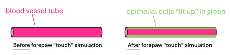
To answer their question, the researchers ran a series of experiments. They measured blood flow, and the calcium in the blood vessels,near active and inactive brain cells. All of the experiments were done in the brains of living, awake mice. Mice are considered “model” organisms. They are useful in research because their bodies function in a very similar way as the human body. For these studies, the researchers used mice that had been genetically altered, called transgenic mice. The mice had been altered so that the endothelial cells in their brains would “light up” in green when calcium was present. The researchers also applied a dye to the blood of the mice so that their blood vessels would “light up” in pink. These methods were used so that the researchers could see both where the blood vessels were (because they would appear pink) and when calcium was released in the endothelial cells of the blood vessel (because it would appear green).
In the first experiment, the researchers wanted to understand how endothelial cells responded when brain cells were active. To activate the brain cells, they simulated touch in the mouse’s paw. When researchers “touched” the paw, they saw that the endothelial cells, which form the walls of the blood vessels, light up in green (Figure 1). This meant that activating brain cells causes nearby endothelial cells to release calcium.
Next, the researchers explored the role of calcium. To do this, they looked at how wide the blood vessel was and counted how many blood cells passed through it before and after calcium was released in the epithelial cells. The researchers found that the blood vessel was relatively narrow before the calcium was released (Figure 2). However, immediately following calcium release, they found that the blood vessel was wider. The researchers also found that the wider blood vessel allowed more blood cells to pass through it, which would allow more nutrients to reach the active brain cells. Based on these results, the scientists concluded that when calcium is released by the endothelial cells, the blood vessels widen to increase blood flow.

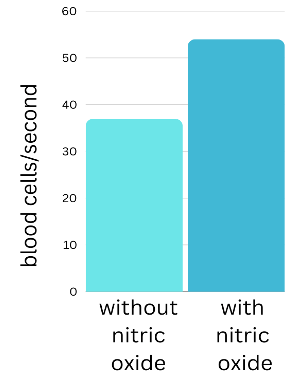
Finally, the researchers wanted to understand why calcium release was leading to wider blood vessels and more blood flow. From previous studies, the researchers already knew two things: (1) that calcium can cause the cell to make a chemical called nitric oxide, and (2) that in other places of the body, nitric oxide causes pericytes to relax, and blood vessels to widen. Based on this knowledge, the researchers predicted that calcium release causes the endothelial cells to make nitric oxide, which then causes the blood vessels to widen. To test this, the researchers added nitric oxide to the brain to see if it caused the blood vessels to widen. The researchers found that more blood cells passed through the blood vessels when nitric oxide was added to the brain (Figure 3). They found that nitric oxide increased blood flow in the brain just like calcium, suggesting that calcium widens blood vessels by causing the body to make nitric oxide.
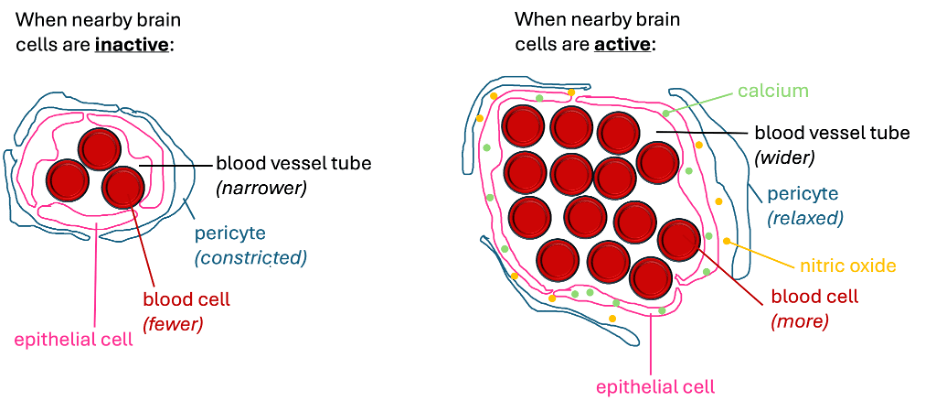
The results of these three experiments allowed the researchers to describe how blood flow is regulated in the brain. They concluded that when the brain cells are active, endothelial cells release calcium. The release of calcium causes the body to make nitric oxide. The nitric oxide then causes the blood vessels to widen, likely by allowing the pericytes to relax. Finally, the widened blood vessels allow more blood cells and nutrients to pass through the tube and reach the active brain cells (Figure 4).
What My Science Looks Like: In these studies, the researchers were able to see real blood vessels, blood flow and calcium release in the brains of living, awake mice. To do this, the researchers had to take several steps. First, because blood vessels and calcium are very small, the researchers had to use a special process called microscopy. Microscopy involves visualizing something very small with a magnification device. Secondly, they needed to be able to tell the difference between calcium and all of the other things inside the brain. Thus, the mice that were used in this study were different from other mice. They had an altered genetic code that allowed their bodies to produce a green fluorescent protein to detect when calcium was higher in their blood vessels. Researchers could shine a light within a particular wavelength on the brain and the protein would fluoresce green, or “light up”, when calcium was elevated. More green light suggests that more calcium is present. When researchers shine a laser at a fluorescent protein, causing it to give off light, and visualize it using a microscope, this process is called fluorescent microscopy.
The research team also used fluorescent microscopy to visualize blood cells and blood vessels. They added a fluorescent dye that sticks to blood cells and blood vessels in the brain. The dye that they used for the blood cells and blood vessels responded to a different wavelength of light than the fluorescent protein used to see calcium. Calcium would fluoresce green, but the blood cells and blood vessels would fluoresce pink. These steps allowed the scientists to see changes in calcium, blood vessel size, and blood flow all at the same time, allowing them to understand how they might influence one another (Figure 5).

The Big Picture: Blood flow is important because our brain cells need energy to function properly. We need to be able to think, act, and experience sensation. When brain cells don’t receive the energy they need, they can become unhealthy. This is something that can happen to humans as we age. As we age, our brains are not as effective at regulating blood flow. Eventually this can lead to dementia. Dementia affects millions of Americans ages 65 and older. Affected individuals begin to forget memories, both from the distant and recent past, and can also develop a change in their personality. It can be very devastating for both the affected individual and their loved ones. If scientists can understand how activity-dependent blood flow in the brain works, they can try to develop treatments, which would improve the quality of life for many aging adults and their families.
Decoding the Language:
Blood cells: Blood cells are the cells in the bloodstream that are responsible for transporting oxygen and nutrients, like vitamins, minerals, sugars, fats, and proteins around the body.
Blood flow: Blood flow refers to the passage of blood cells, and other components of the blood, through the blood vessels.
Blood vessels: Blood vessels area network of tubules through which the bloodstream travels from place to place in the body.
Brain cells: Brain cells are cells within the brain that communicate with one another through both electrical and chemical signaling. Brain cells produce thought, action, and/or sensation (smell, touch, hearing, taste, and sight). The brain contains billions of cells, so at any given moment, some are actively signaling, and some are not, depending on what you are thinking, doing, or sensing.
Calcium: Calcium is a mineralfound mainly in dairy products and is needed for healthy teeth, bones, and other issues. It plays many roles in making sure our bodies function properly; not just keeping your bones strong!
Cross-section: A cross-section isthe view that emerges if you were to cut straight through something to expose the inside.
Dementia: Dementia is a condition characterized by the loss of memory and thinking skills, such as problem-solving and making judgements, and often leads to personality changes.
Endothelial cells: Endothelial cells that form the “walls” of the blood vessels.
Fluorescent microscopy: Fluorescent microscopy is a technique that uses a microscope that can shine various wavelengths of light onto a fluorescent sample to view it. Researchers will shine a light on a protein or molecule with the fluorescent properties, and the sample will emit the fluorescent light. The light will then be filtered through the microscope to create an enlarged image that shows the sample. This technique is used to view really small samples that normally cannot be seen by the human eye.
Fluorescent protein: A fluorescent protein is often designed by people to have a special property that allows it to give off light when exposed to a specific wavelength of light.
Genetic code: DNA contains a pattern of instructions, through genes, that essentially tell the body how to build itself. Just like a construction worker needs to know how to arrange the boards, your body needs to know how to organize all of its building blocks. The genetic code is (metaphorically) comparable to a construction worker’s blueprint.
Microscopy: Microscopy is a scientific techniqueused to magnify things that normally cannot be seen by the human eye unaided.
Nitric oxide: Nitric oxide is a chemical compound made up of two chemical elements, nitrogen and oxygen, that are bound together. It plays many important roles inside of the body and takes the form of a gas (rather than a liquid or solid), which allows it to spread quickly and easily.
Nutrients: Nutrients are derived from a nutritious diet, and include vitamins, minerals, fats, sugars, and proteins. They travel through the bloodstream, on blood cells, to reach all cells of the body and to keep them, and therefore you, healthy and functioning properly.
Pericytes: Pericytes arecells that wrap around blood vessels, to “squeeze” or “relax”, causing the blood vessel to shrink/constrict or widen, respectively.
Postdoctoral: The stage of scientific training that many researchers take after earning a doctoral degree (PhD). Many researchers work as a postdoctoral researcher for several years before running their own research laboratory.
Predict: The term “prediction” is often used in science to refer to what the researchers expect the data to look like, based on prior knowledge.
Transgenic mice: Transgenic mice are mice with altered genetic codes. Researchers will alter the genetic codes of mice to help them better understand how biological systems work.
Wavelength: A wavelength is a property of light that, when varied, makes it appear different colors.
Learn More:
An article from GiveBlood about the role of blood flow in the body.
An article from the University of London on how the brain uses energy.
An article from the Scientific American about why the brain needs energy.
An article from Columbia University Irving Medical Center about the prevalence of dementia.
An article from the National Institute on Aging about how Alzheimer’s disease affects the brain.
Synopsis edited by Dr. Rosario Marroquin-Flores (she/her), Assistant Professor, James Madison University.
Download this article here
